Doctoral researcher in experimental soft-matter and biological physics at Saarland University. I study the mechanics, flow and aggregation of red blood cells, from single cells to dense suspensions, using in vivo microscopy, image analysis and physical modelling.
Since 2022 I have been working towards a doctorate (Dr. rer. nat.) in the group of Prof. Christian Wagner at Saarland University. My Master's thesis dealt with the sedimentation of erythrocytes in colloidal-silica density gradients. My current work covers detection and tracking pipelines for microscopy data, 2D/3D image segmentation, shape analysis, and models of how red cells move, deform and aggregate.
The position is funded by a Young Investigator Grant awarded to Dr. Alexis Darras. In 2025 the work received the Best Poster Award at the Annual European Rheology Conference (AERC) in Lyon.
MATLABPythonC++ / CUDAImage analysisEmbedded systems
Collaborating institutions
Saarland UniversityChristian Wagner, Lars Kaestner, Thomas John, Kirill Korneev, Yazdan Rashidi, Khadija Larhrissi, Min Qiao, Diana Örüm, Nikolas Lerch, Luca Hastenteufel
KU LeuvenErin Koos, Pavlik Lettinga, Christoph Haessig
University of ZürichAnna Bogdanova, Asya Makhro
University of PaviaGiampaolo Minetti
Université Grenoble AlpesChaouqi Misbah
University of GöttingenCatalina Posada, Michael Rostas
Funding
Young Investigator Grant of Saarland University (A. Darras) · DFG research unit FOR 2688 · Research Foundation – Flanders (FWO), project 1108426N.
02 · Research
Selected projects
Figures are taken from the respective studies and talks; references are given with each project. Click a figure to enlarge it.
PlasmaFITC-dextranHealthy red cellsCellTracker Deep RedRigidified cellsPKH26Mergedall channels
Intravital fluorescence microscopy in a dorsal skinfold chamber: the three labels are recorded in separate channels and tracked frame by frame.
i.
Blood cells in vivo: tracking at vessel bifurcations
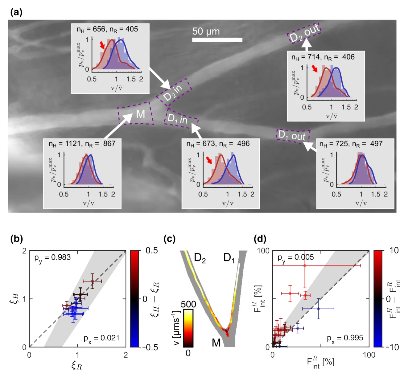
Detection and tracking pipelines for intravital microscopy. White blood cells are classified as rolling or flowing along the vessel wall and followed through bifurcations. In a second study, healthy and artificially rigidified red cells are compared at vessel junctions: rigid cells spend less time at the branch apex, which changes how blood is partitioned between the daughter vessels. This is relevant to diseases with stiffened red cells, such as malaria and sickle-cell disease. My contribution: video processing, tracking pipeline, data analysis, statistics and figures.
White blood cells: ongoing investigations with Khadija Larhrissi, AG Wagner. Rigid versus healthy red cells: Rashidi*, Maurer*, Wrublewsky*, Larhrissi, John, Gidley, Toye, Kaestner, Wagner, Laschke, Darras. Biophysical Journal (2026). doi:10.1016/j.bpj.2026.03.023
Velocity distributions of healthy (H) and rigid (R) cells along a bifurcation (a) and the resulting statistics (b–d).
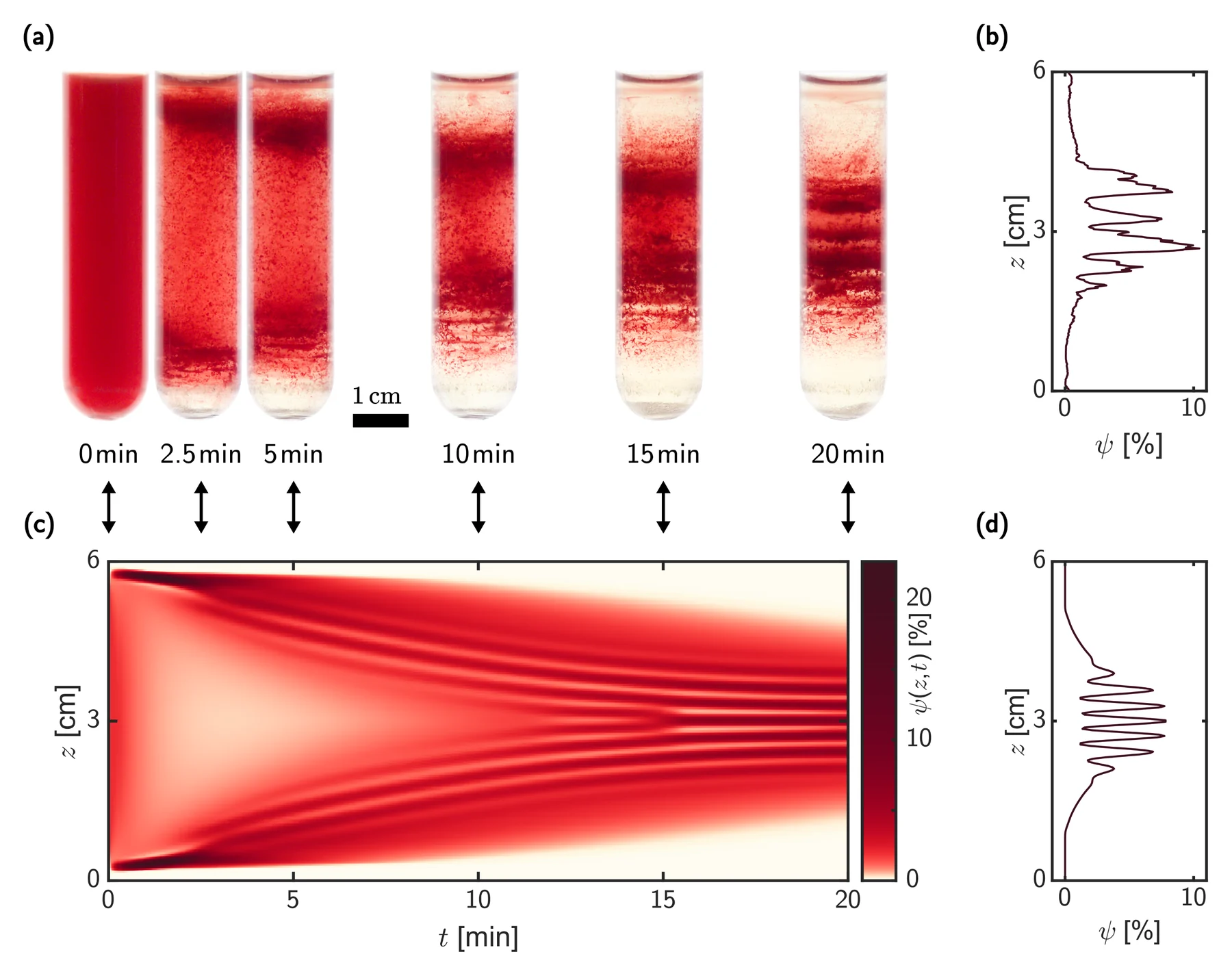
Band formation over 20 minutes: tube photographs (a) and measured concentration profile (b), compared with the space–time evolution (c) and final profile (d) of the continuity model.
ii.
Band patterns in density gradients
Centrifuged red cells settle into discrete bands, which were long interpreted as evidence of discontinuous cell ageing. Using a continuity equation that includes aggregation, we showed that the bands result from a competition between cell aggregation and net buoyancy. Related work explains the bands in classic Percoll gradients and the resulting limits of age separation.
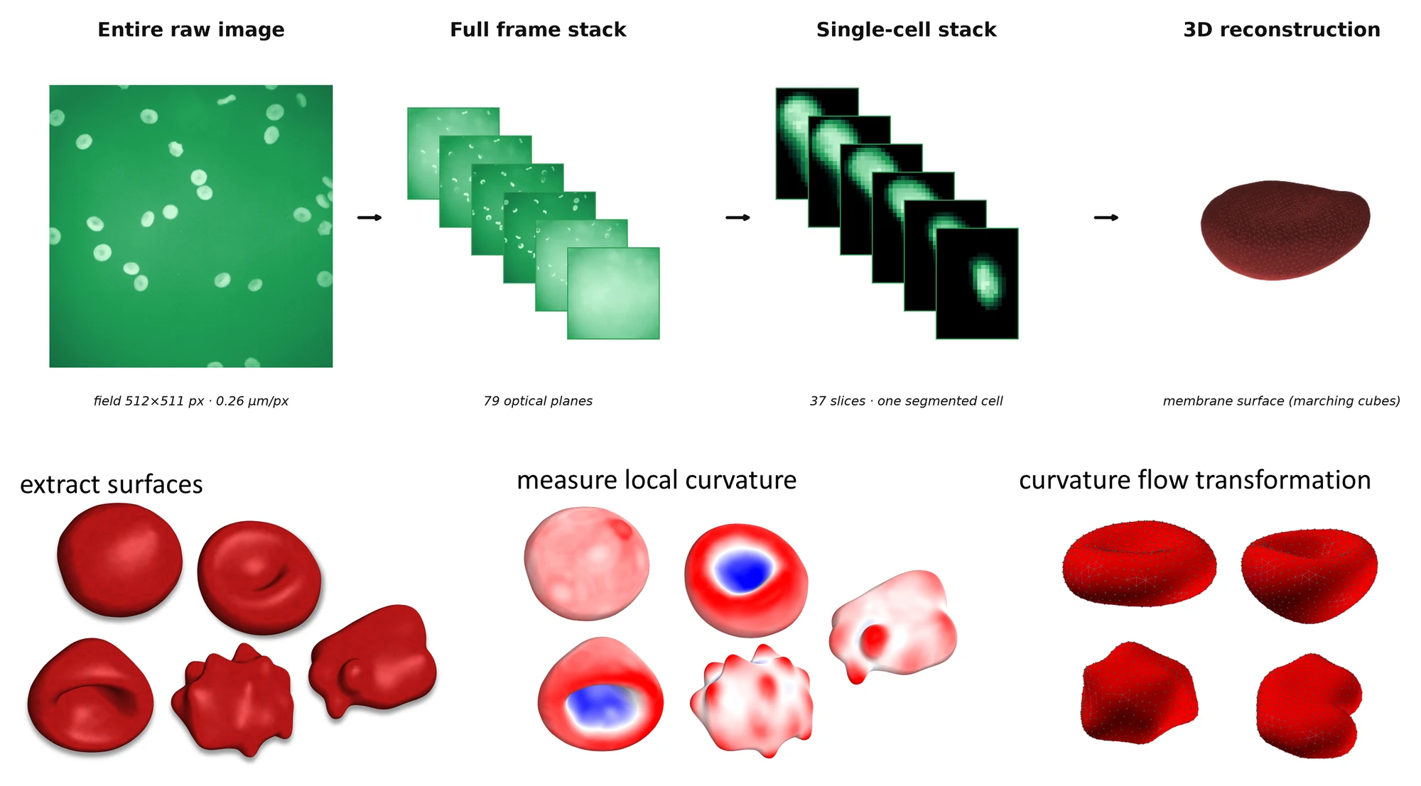
From the confocal stack to the cell surface: segmentation and 3D reconstruction (top), surface extraction, local curvature and Willmore curvature flow (bottom).
iii.
3D phenotyping of red-cell shape
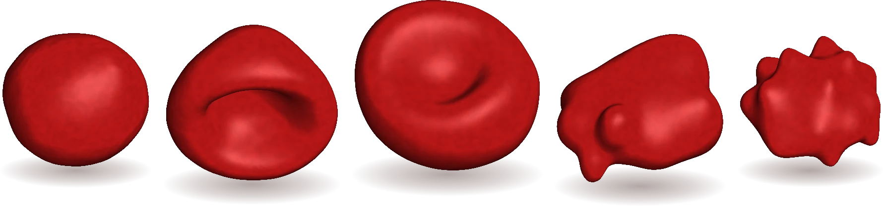
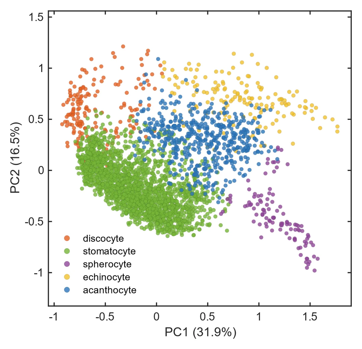
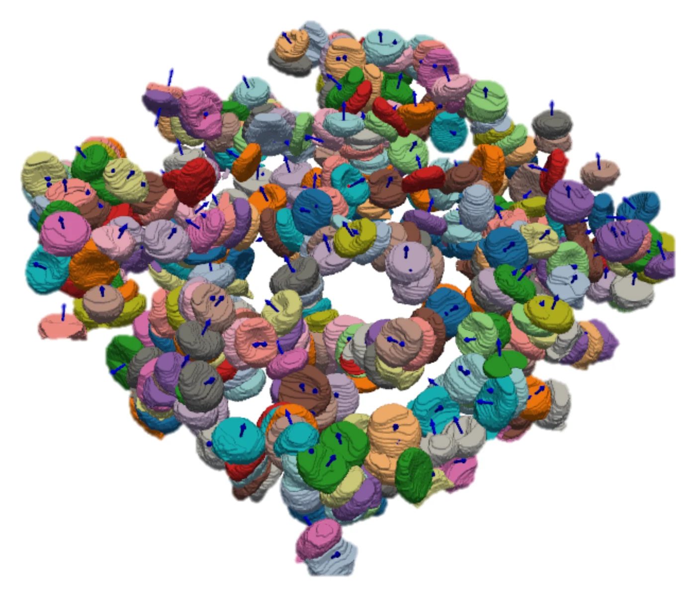
Cell surfaces from confocal and STED stacks are smoothed by Willmore curvature flow and decomposed into spherical harmonics, which places each cell on the stomatocyte–discocyte–echinocyte axis. A random-forest classifier reaches 92% accuracy on 730 cells and separates shape changes that are difficult to distinguish by eye. Developed together with Nikolas Lerch.
Building on the 3D red-cell phenotyping of Simionato, Quint et al. AG Wagner, Saarland University.
Shape classes in the plane of the first two principal components.
Further projects
Cytosol viscosity and density
Buoyant-density centrifugation gives the density distribution of red cells and, via the relation between cytosol viscosity and density, their intracellular viscosity. The results provide single-cell parameters for blood-flow models. My contribution: density-gradient preparation and measurements.
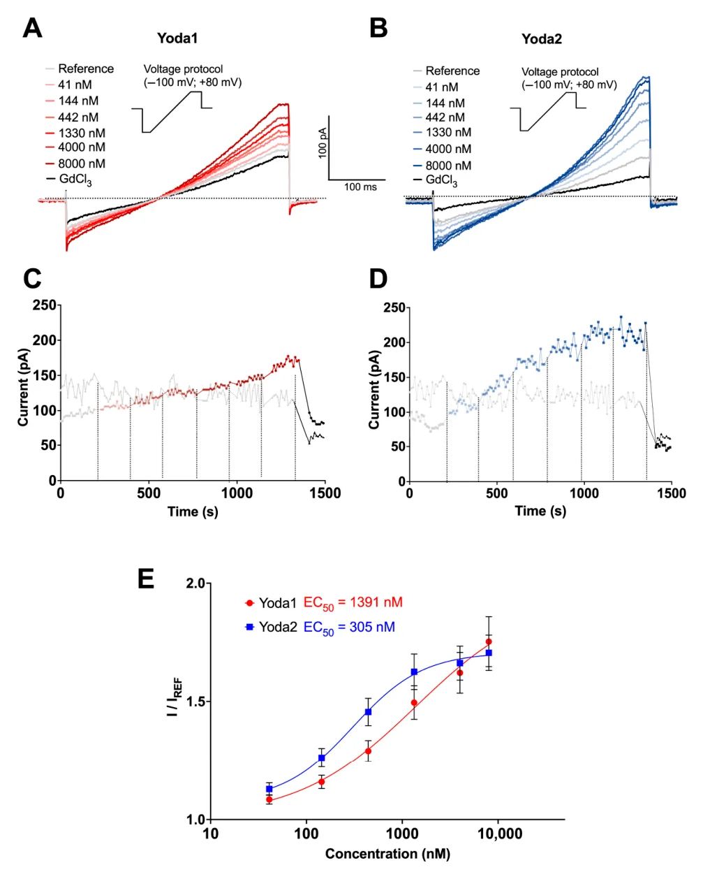
Automated patch clamp and calcium imaging are used to compare how the chemical activators Yoda1 and Yoda2 open the mechanosensitive Piezo1 channel in red cells.
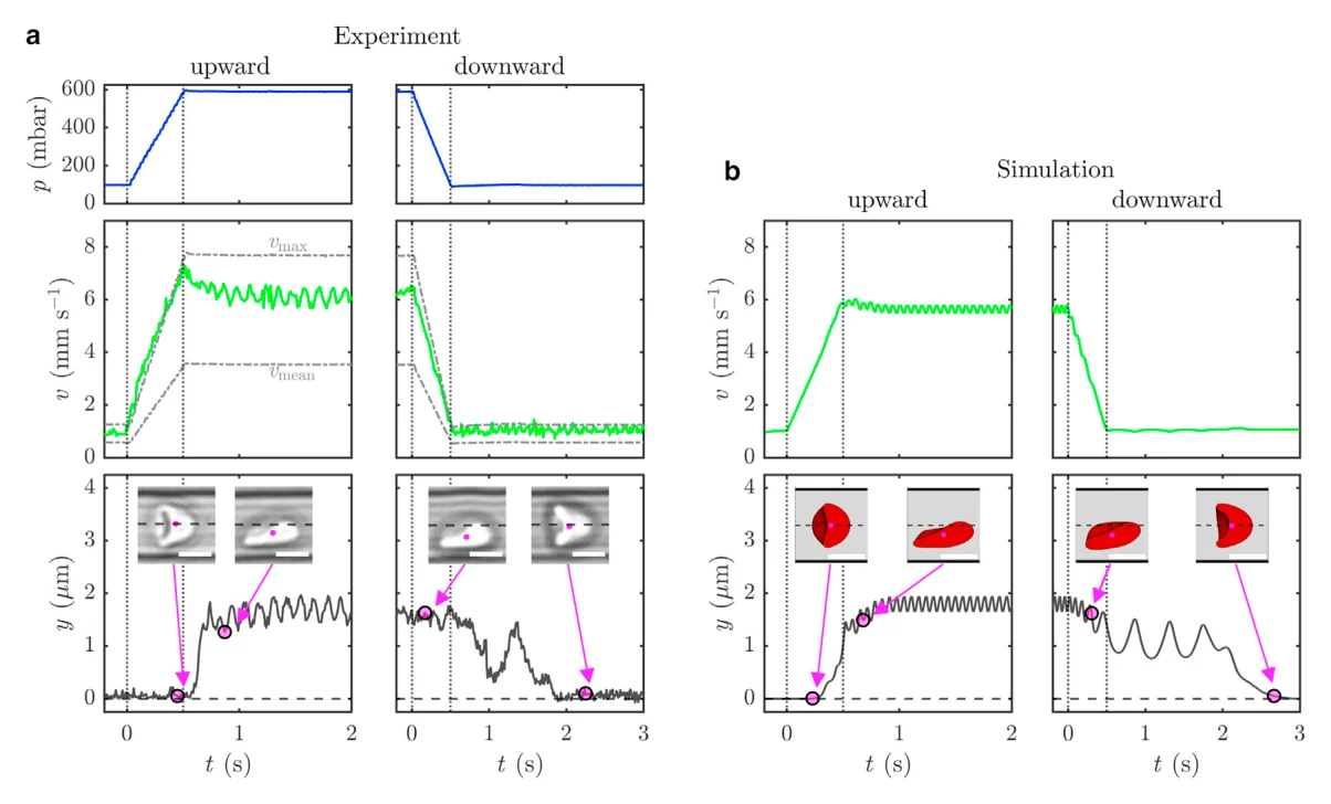
In capillary flow, red cells adopt either a symmetric croissant or an off-centred slipper shape. Under time-dependent flow, the croissant-to-slipper transition is considerably faster than the reverse and depends on the flow ramp rate. Cells were tracked in a co-moving frame using a custom PID-controlled microscope stage.
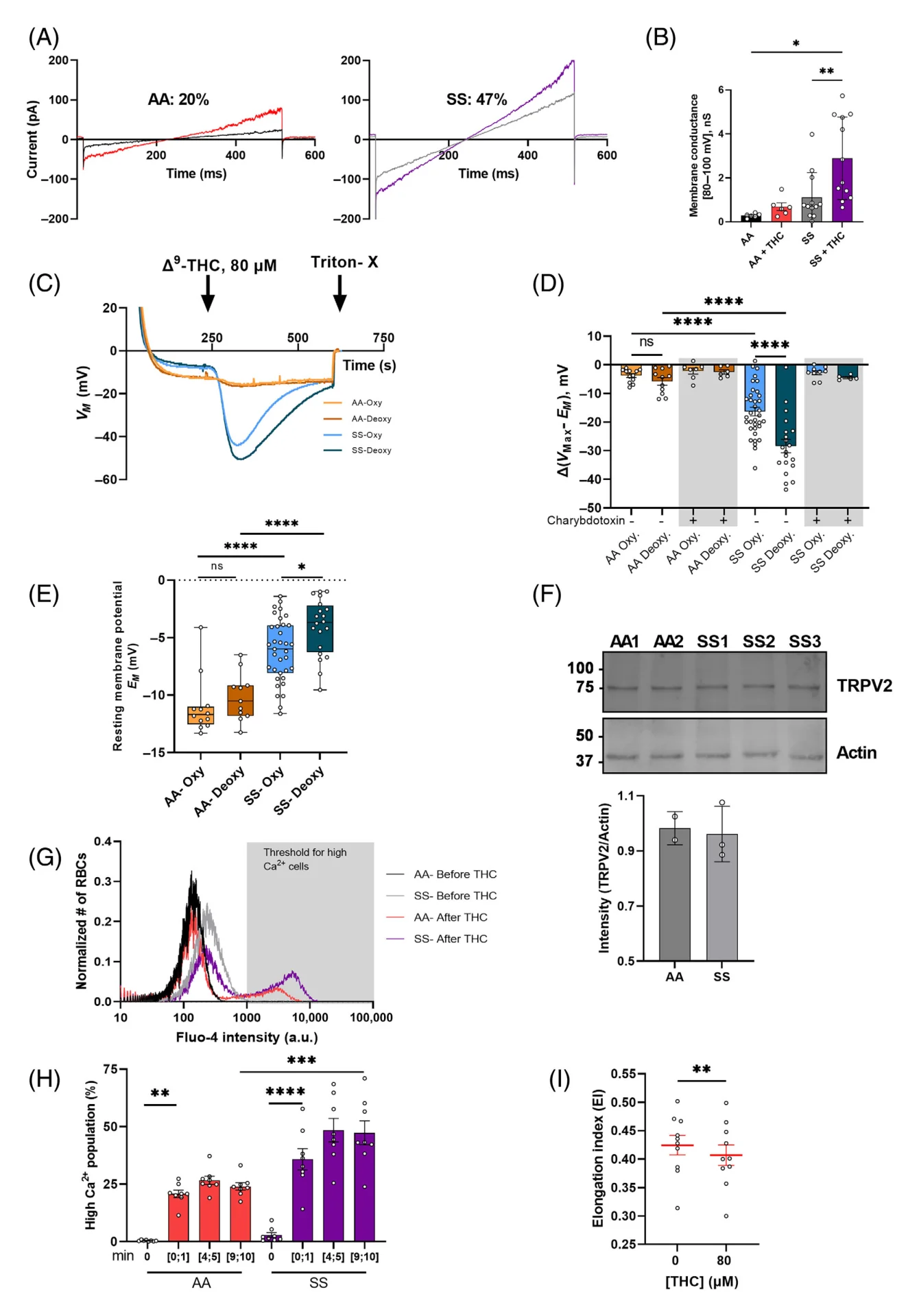
Δ⁹-THC activates cation channels in red cells, which raises intracellular calcium and leads to dehydration and shape change. After THC exposure, a large fraction of sickle cells shifts into the high-calcium range, suggesting that cannabis use may be harmful in sickle-cell disease.
Hatem*, Esperti*, Murciano*, Qiao*, … Maurer, … Egée. American Journal of Hematology 98(12) (2023). doi:10.1002/ajh.27109
Deposits of drying blood droplets
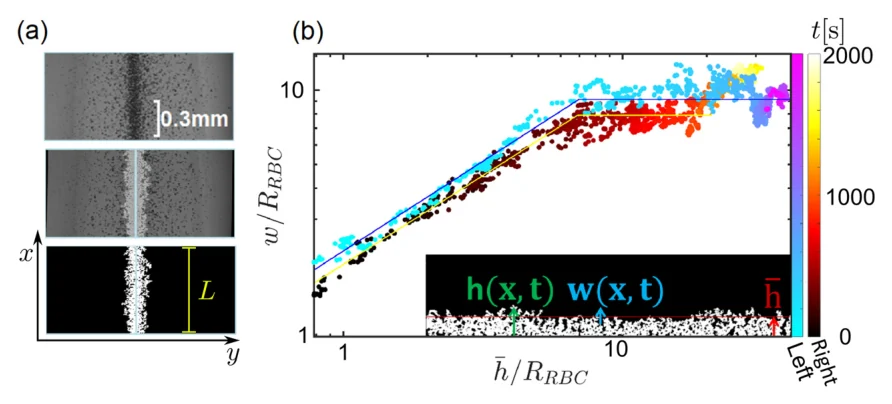
Drying droplets usually leave a ring-shaped deposit (coffee-ring effect). At low red-cell concentration, the deposit instead grows from a central edge. This growth is quantified via interface growth and roughness scaling, with potential use in blood-drop diagnostics.
Ongoing work: optical-tweezers measurements of the attraction between two cells are used to derive a pair-interaction potential, which enters a percolation model of aggregated blood as a gel.
Ongoing, with K. Korneev (AG Wagner) and C. Haessig (KU Leuven). 3D reconstruction and recording of the cells shown by C. Haessig.
Responsible for content: Felix Maurer, Campus E2 6, 66123 Saarbrücken, Germany. Telephone +49 (0)681 302 - 2977, email mail@felixmilanmaurer.com.
External links: despite careful content control we assume no liability for the content of external links; the operators of the linked pages are solely responsible for their content.
Messages sent with the form (name, email address, optional subject, message) are used only to answer your request. They are forwarded by email to mail@felixmilanmaurer.com and stored on this website's server, hosted by Alfahosting GmbH, Halle (Saale), Germany, for at most 12 months, then deleted automatically. Legal basis: Art. 6(1)(b) and (f) GDPR. You can request information about or deletion of your data at any time via the email address above.